Up to 40% of patients with breast cancer present with metastases in sentinel lymph nodes at diagnosis. Sentinel lymph nodes are the initial sites where breast cancer cells might spread. Axillary lymph nodes are secondary metastasis sites. Lymph nodes are important hubs for immune cell activation (‘priming’), where naïve B and T cells, the primary workhorses of the immune system, recognise and are activated against their specific targets, thereby enabling them to initiate an effective immune response. However, in metastatic lymph nodes immune responses are often skewed towards immune suppression allowing for further tumour progression — a process that immunotherapy treatments seek to reverse.
In a new Research Article in Molecular Oncology, Rye et al. [1] mapped the phenotype, indicative of activation status, and distribution of immune cells in non-metastatic and metastatic sentinel lymph nodes and in metastatic axillary lymph nodes from 52 breast cancer patients. Having a better understanding of immune profiles over the course of early lymph node metastasis could enable the assessment of novel immunotherapy treatments for metastatic breast cancer.
T cell dynamics at lymph node sites reflect tumour dissemination
Rye et al. collected lymph node tissues representing different stages of tumour progression from breast cancer patients and analysed single cells based on proteins expressed on their cell surface.
Both metastatic and non-metastatic sentinel lymph node samples displayed a roughly equal proportion of naïve- and memory CD4+ and CD8+ T -cells. In contrast, metastatic axillary lymph node samples had a greater abundance of memory CD4+ and CD8+ T cells than naïve ones. Additionally, authors showed a positive correlation between a higher tumour burden in axillary lymph nodes and the numbers of CD8+ T cells and regulatory T cells (T regs). T regs typically suppress overt immune responses to ensure homeostasis.
T cell exhaustion reflects tumour cell spread
Furthermore, CD4+ and CD8+ T cells as well as T regs in metastatic axillary lymph node samples showed an increased expression of T cell exhaustion markers such as TIGIT and PD-1, compared to metastatic and non-metastatic sentinel lymph node samples. TIGIT and PD-1 act as negative regulators of T cell activation and have been associated with immune evasion in cancer. This suggested that T cell ability to elicit an immune response is inhibited in metastatic axillary lymph nodes.

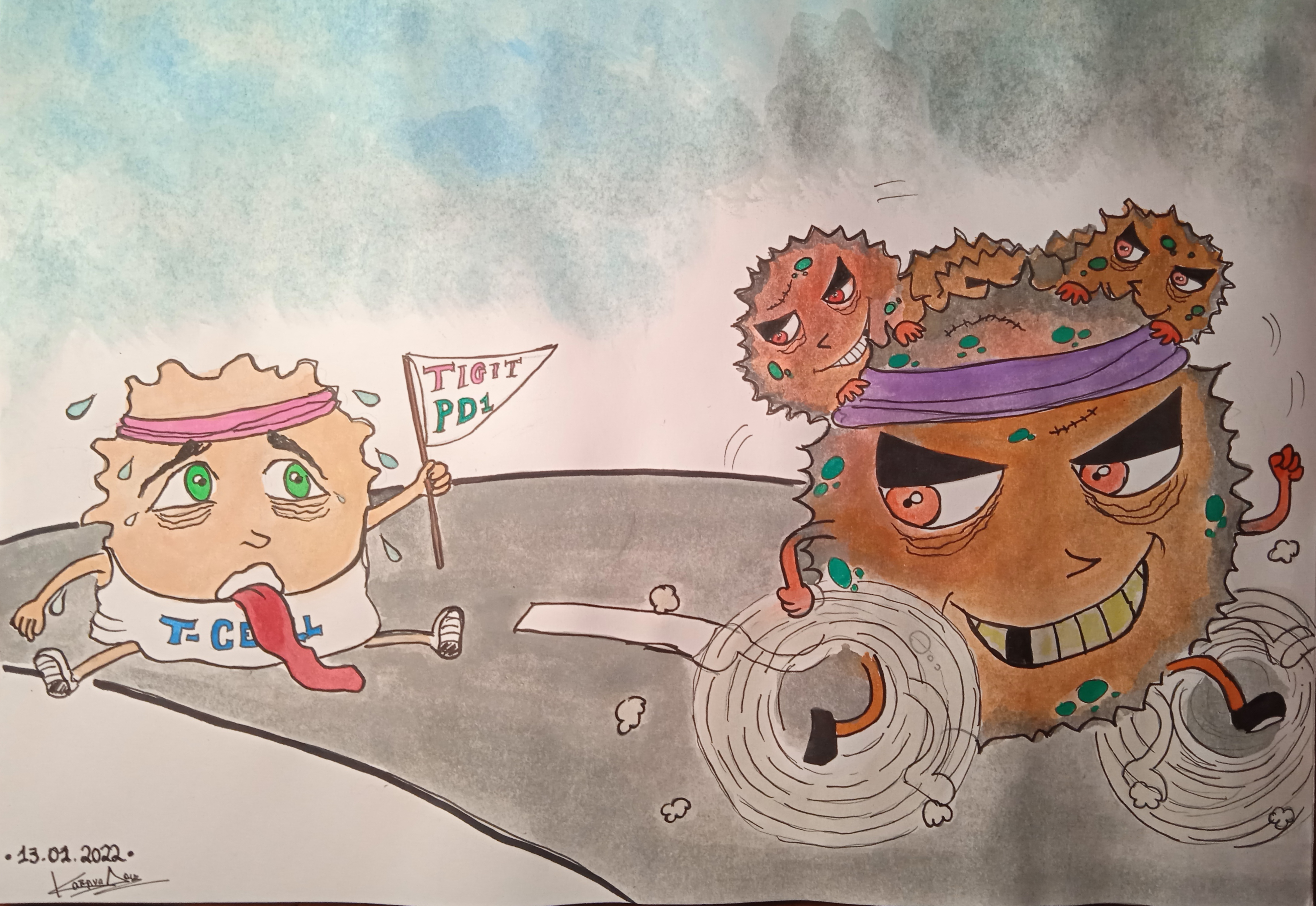
Cartoon representation of an ‘exhausted’ T cell expressing TIGIT and PD1 (T cell exhaustion markers), unable to mount an immune response, resulting in escaping cancer cells. Illustration by Dr Aikaterini Douka (@kateRiNA_douka1).
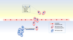
Moreover, a positive correlation was estimated between TIGIT-expressing T cells and increasing tumour burden, implying metastatic cancer cells may directly lead to T cell exhaustion. In addition, the ratio of CD4+ to CD8+ cells in tissue sections stained for tumour and immune cell markers was lower than one in the area of the tissue where the tumour was found and higher than one outside of the tumour area, suggesting skewing towards a CD8+ T cell expansion. What’s more, the presence of T regs was observed in close proximity to CD8+ T cells.
Finally, T cells expressing the TIGIT marker appeared to have dysfunctional T cell signalling, based on reduced ERK phosphorylation. Collectively, the findings of Rye et al. suggest that in areas where cancer cells and immune cells can interact, the T cell distribution is shifted towards exhausted CD8+ T cells and T regs, potentially suppressing tumour immunity altogether.
References
- Rye, I. H., Huse, K., Josefsson, S. E., Kildal, W., Danielsen, H. E., Schlichting, E., Garred, Ø., Riis, M. L., Osbreac, Lingjærde, O. C., Myklebust, J. H. & Russnes, H. G. (2022) Breast cancer metastasis: immune profiling of lymph nodes reveals exhaustion of effector T cells and immunosuppression. Molecular Oncology. 16, 88-103. https://doi.org/10.1002/1878-0261.13047
Molecular Oncology is an Open Access international journal that highlights new discoveries, approaches, as well as technical developments, in basic, clinical and discovery-driven translational cancer research.


Join the FEBS Network today
Joining the FEBS Network’s molecular life sciences community enables you to access special content on the site, present your profile, 'follow' contributors, 'comment' on and 'like' content, post your own content, and set up a tailored email digest for updates.